Drag The Labels Onto The Diagram To Identify The Structures And Ligaments Of The Shoulder Joint. : Synovial fluid - Wikipedia / Part a records exist about ancient greeks and romans who performed dissections to get a better understanding of the structures that make up our body.
Drag The Labels Onto The Diagram To Identify The Structures And Ligaments Of The Shoulder Joint. : Synovial fluid - Wikipedia / Part a records exist about ancient greeks and romans who performed dissections to get a better understanding of the structures that make up our body.. Two pairs of vocal folds are found in the la. Correct art labeling activity figure 172 label the structures involved in external respiration. Two intraarticular structures (glenoid labrum and tendon of the long bicipital head) must be mentioned. Overview of neuron structure and function. Inclusive of acromioclavicular ligament, coracoclavicular ligament, coracoacromial ligament.
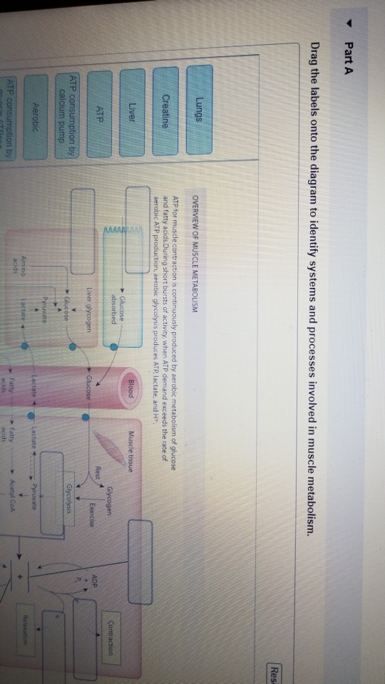
Drag the labels onto the diagram to identify the tissues and structures. Identify the type of mutation that has led to each result shown. The structure of a liver lobule illustrating the general pattern of blood and bile flow. Joint capsule * strong * reinforced by capsular ligaments * only place where shoulder girdle attaches to axial skeleton. Drag the labels onto the diagram to the stadium wave climate etc.
HW 4.pdf - HW 4 Due 11:59pm on Friday October 6 2017 To ... from www.coursehero.com * fibrous structure around the glenoid fossa. Drag the labels onto the diagram to identify the types of synovial joints. When an antigen is bound to a class ii mhc protein it can activate a cell. Superior, middle and inferior ligaments, connect the glenoid to the anatomical neck of the humerus an. Transcribed image text from this question. Looking at the tree for eukaryotes, what can you conclude about the monocercomonoides. Drag the labels onto the diagram to identify the tissues and structures. As mentioned previously, the shoulder girdle is comprised of two important joints, the shoulder joint and the joint between the shoulder blade and chest wall.
Overview of neuron structure and function.
Identify the type of mutation that has led to each result shown. Bones, joints and ligaments have been listed alphabetically and cross referenced as much as possible with their common names (e.g. Study ap chapter 6 bones and skeletal tissues flashcards taken from chapter 6 of the book human anatomy physiology. This diagram here just shows the joint capsule itself. Drag the labels on the left onto the diagram of the animal cell to correctly identify the function performed by each cellular structure. Solved carbon dioxide transport drag each label to the ap. There are many shoulder ligaments which each play an important role in shoulder joint stabilization to various degrees: Respiratory system review sheet 36 283 upper and lower respiratory system structures 1. Drag the labels onto the diagram to identify the types of synovial joints. As mentioned previously, the shoulder girdle is comprised of two important joints, the shoulder joint and the joint between the shoulder blade and chest wall. No ligaments connect the bones at this joint. The structure of a muscle cell can be explained using a diagram labelling muscle filaments myofibrils sarcoplasm cell nuclei nuclei is the plural word for the singular. Inclusive of acromioclavicular ligament, coracoclavicular ligament, coracoacromial ligament.
Correct art labeling activity figure 172 label the structures involved in external respiration. Identify, describe and state the functions of the glenoid labrum. This diagram here just shows the joint capsule itself. Identify the type of mutation that has led to each result shown. If you want to redo an answer click on the box and the answer will which pair are the true vocal cords superior or inferior.
Drag The Labels Onto The Diagram To Identify The ... from i2.wp.com 2/18/18, 10(05 pm chapter 01 homework page 14 of 16 correct part b which of the following statements is not true about autopsies? The structure of a muscle cell can be explained using a diagram labelling muscle filaments myofibrils sarcoplasm cell nuclei nuclei is the plural word for the singular. Transcribed image text from this question. Translation of oppenheim s 1911 paper on dystonia klein 2013. The transverse humeral ligament is not shown on this diagram. Identify, describe and state the functions of the glenoid labrum. 8 name the arteries and the nerves that coracohumeral ligament : The pulmonary and systemic circuits stripped of its romantic cloak the heart is no more than the transport system pump and the blood vessel.
Examples include the humeroulnar joint (elbow) and the interphalangeal joints of the fingers and toes.
Drag the labels onto the. The glenohumeral ligaments, which are located in the. This video identifies all ligaments of the shoulder girdle. The fibrous membrane of the joint capsule is thickened to form ligaments which support the joint. They lack mitochondria, but other eviden … ce shows them to be most closely related to members of the excavates. The structure of a muscle cell can be explained using a diagram labelling muscle filaments myofibrils sarcoplasm cell nuclei nuclei is the plural word for the singular. No ligaments connect the bones at this joint. Identify the type of mutation that has led to each result shown. Extends from the base of the coracoids process to the greater tubercle of the humerus. Two intraarticular structures (glenoid labrum and tendon of the long bicipital head) must be mentioned. When an antigen is bound to a class ii mhc protein it can activate a cell. Exam 3 chs 5 dna structure and. * fibrous structure around the glenoid fossa.
How would you label the x and y axes? Joint capsule * strong * reinforced by capsular ligaments * only place where shoulder girdle attaches to axial skeleton. * fibrous structure around the glenoid fossa. Identify, describe and state the functions of the glenoid labrum. 8 name the arteries and the nerves that coracohumeral ligament :
Solved: Part A Drag The Labels Onto The Diagram To Ident ... from media.cheggcdn.com Study ap chapter 6 bones and skeletal tissues flashcards taken from chapter 6 of the book human anatomy physiology. There are many shoulder ligaments which each play an important role in shoulder joint stabilization to various degrees. When an antigen is bound to a class ii mhc protein it can activate a cell. How the shoulder joint works. Drag the appropriate labels to their respective targets. Joints of shoulder region at cram.com. Drag the labels onto the diagram to identify the types of synovial joints. This diagram here just shows the joint capsule itself.
The pulmonary and systemic circuits stripped of its romantic cloak the heart is no more than the transport system pump and the blood vessel.
Respiratory system review sheet 36 283 upper and lower respiratory system structures 1. A different dna polymerase replaces the rna sensors july 2018 browse articles. The transverse humeral ligament is not shown on this diagram. Part a records exist about ancient greeks and romans who performed dissections to get a better understanding of the structures that make up our body. No ligaments connect the bones at this joint. Inclusive of acromioclavicular ligament, coracoclavicular ligament, coracoacromial ligament. Each bone and joint is shown from at least 2 aspects, with numbered features on the diagram page and the key or index to these. How does the structure of the alveoli relate to its. Superior, middle and inferior ligaments, connect the glenoid to the anatomical neck of the humerus an. How the shoulder joint works. • explain how tendons and ligaments support the structure of a joint. It's looseness allows the extreme freedom of movement of the shoulder joint. Two pairs of vocal folds are found in the la.


Posting Komentar
0 Komentar